
Confocal laser scanning microscope
-
Inverted confocal with temperature and CO2 control
- Objectives: 10x, immersion: 20x 63x water, 40x 100x oil
-
405 nm violet excitation
-
line, frame and stack sequential scanning
-
Spectrometric imaging with five freely selectable emission ranges and settings for any fluorochrome
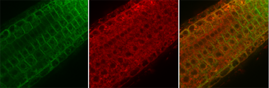
Arabidopsis root showing colocalisation of a membrane protein and a ER marker (Bangjun Wang, Markus Geisler, Alexander Schulz, unpubl).
|
Laser lines |
Beam splitter |
Emission |
Detectors |
|
405 nm UV,V |
freely tuneable AOBS |
freely selectable range |
5 PMTs |
|
458, 476, 488, 496, 514 nm |
|||
|
543, 633 nm |
Details
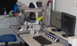 Microscope
Inverted Point Scanning Confocal SP5 II, Leica Microsystems
Location
North Campus, CAB, August Krogh Building, room 01-6-637B
Typical applications
Live imaging of cells and tissues, (co-) localisation of fluorochromes and fluorescent proteins, FRET, FRAP
Consultation, training and problem reporting
Nynne Meyn Christensen
Booking
Online via PPMS.