
Robot microscope
-
Spinning disk confocal imaging in plates or slide format
-
Environmental control: temperature, CO2, humidity
-
Objectives: 5x, 20x, and 20x and 60x water immersion
- 5 laser lines 375 nm, 425 nm, 488 nm, 561 nm and 640 nm
-
Two sCMOS image sensors
- Emission filters: 435-480, 435-550, 570 - 630, 650 - 760, 435 - 515, 465 - 530, 571-596, 605-630, 500 - 550, 500 - 530, 515 - 550, 650 - 680 and 690 - 720
- Software guides to optimal setting for fluorophores
- Prescan - rescan with AI-powered target detection allows for scan of whole sample area, detection of relevant sample parts and rescan with higher magnification
-
Dispenser unit for addition of compounds to wells/slide
-
Laser based auto focus
-
Script-based Image and Data analysis program that can run during the experiment
Details
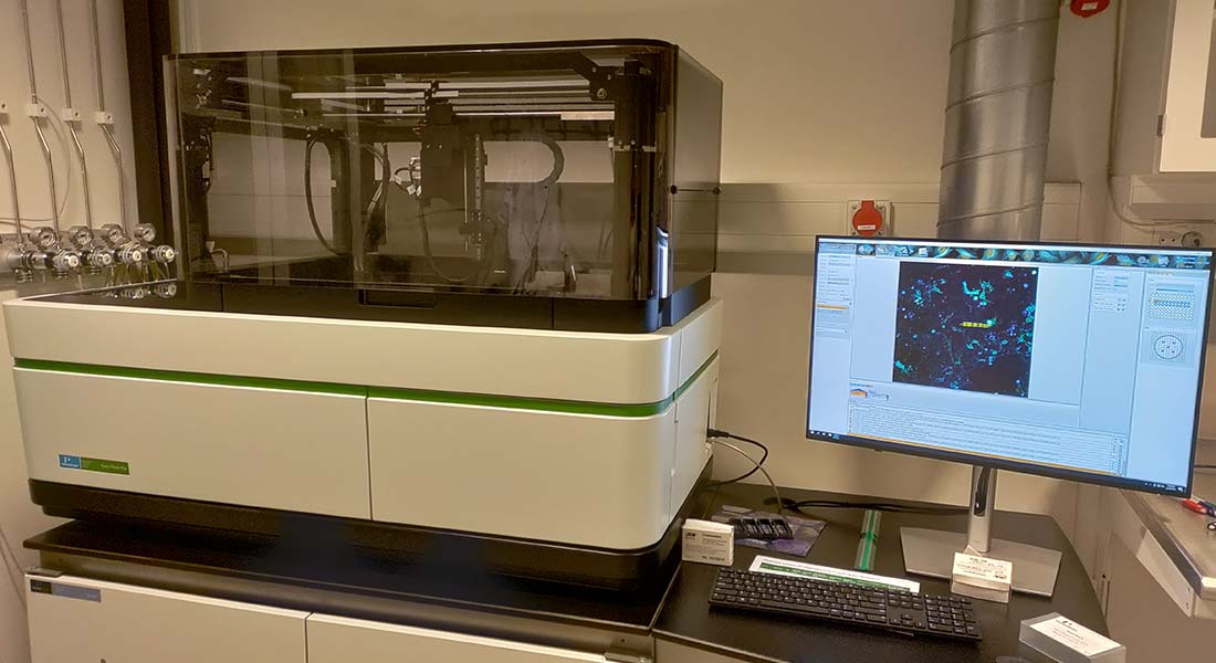
Microscope
High-content screening spinning disc confocal microscope, Opera Phenix, PerkinElmer
Location
North Campus, August Krogh Building,
Universitetsparken 13, 6. floor, room 644.
Typical applications
High throughput live imaging, library screening
Consultation
Nynne Meyn Christensen
Michael Lisby
Training and problem reporting
Booking
Online via PPM.