
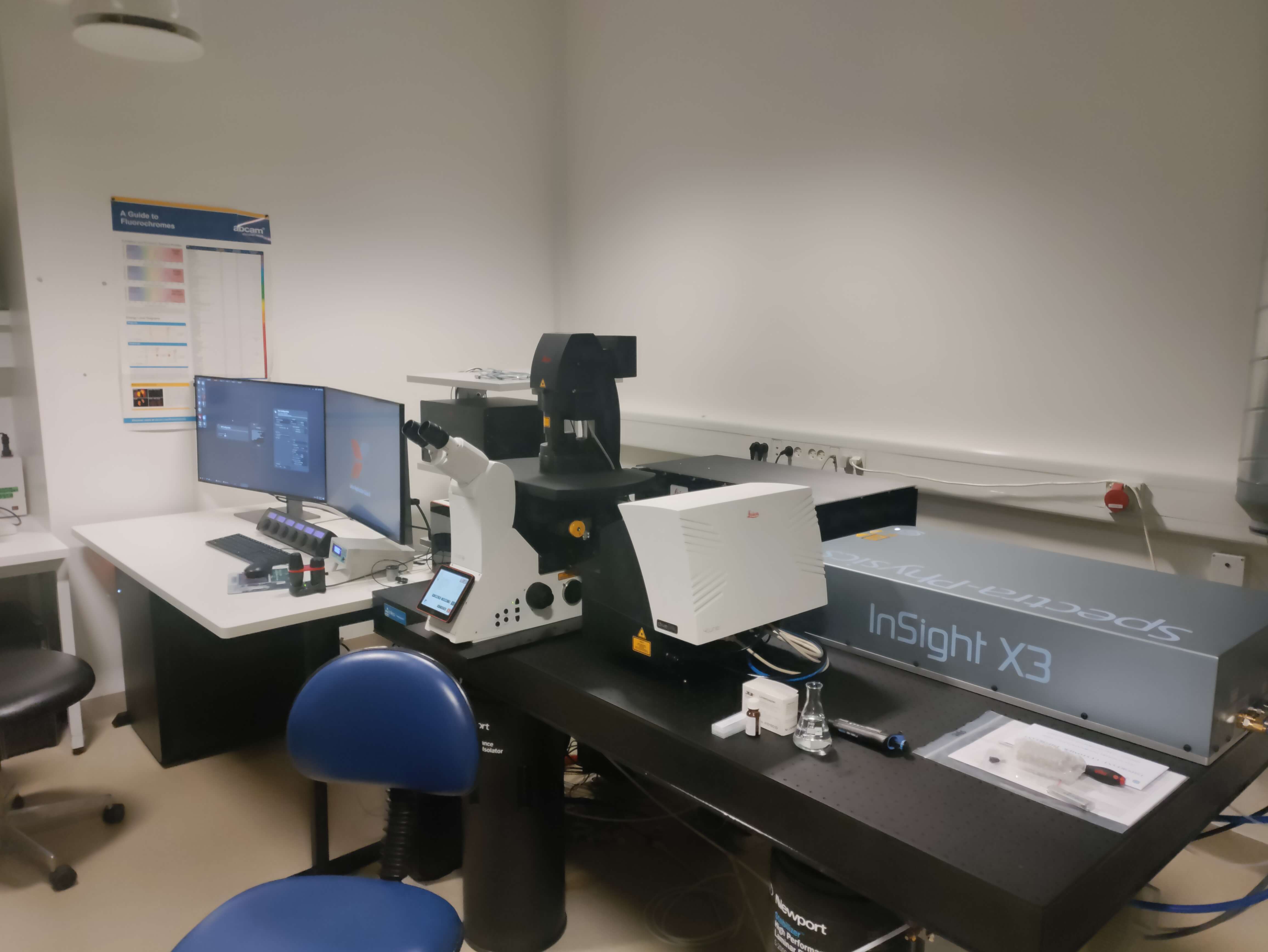
Confocal, Multiphoton, FLIM microscope
- Inverted point scanning confocal microscope with galvo scanner and navigator software
- Objectives: 20x (0.75) dry, 25X (0.95) water, 40 (1.10) water, 40X (1.2) Glycerol.
-
Supercontinuum white light laser (WLL) with up to 8 excitation lines between 440nm and 790nm
-
Two Multiphoton (MP) lasers: 680 – 1300 nm + fixed laser at 1045 nm
-
3 internal HyD detectors with freely tunable filters
-
2 non-descanned detectors with freely tunable filters
-
MP and WLL FLIM on 2 internal HyD and 2 non-scan detectors and tunable filters
-
FLIM software including phasor plot.
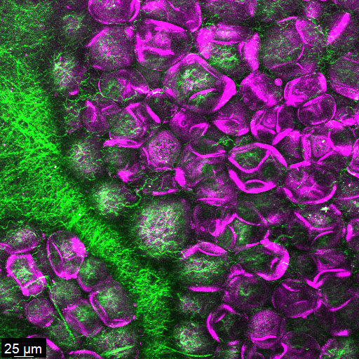
Label-free imaging of mouse kidney with multiphoton laser. Green: collagen (SHG) and purple: Membranes (THG)
Details
Microscope
Leica Stellaris 8 DIVE-FALCON microscope. Point-scanning Confocal and Multiphoton Flourescence liftetime imaging microscope.
Location
North Campus, August Krogh Building,
Universitetsparken 13, 6. Floor, room 644
Typical applications
Live imaging with environmental control. f cells, tissue and organs, label-free imaging (SHG, THG) protein localization, intracellular ion concentrations, FRAP, FRET. Video-rate FLIM with phasor plot analysis
Consultation
Nynne Meyn Christensen
Jakob Grunnet Knudsen
Training and problem reporting
Booking
online via PPMS.