
Upright CLSM
Confocal laser scanning microscope
-
Confocal imaging with up to 25 fps
- Objectives: 10x, immersion: 20x 63x water, 40x 63x oil; dipping: 20x 40x 63x water
-
Supercontinuum white light laser (WLL) with up to 8 excitation lines between 470nm and 670nm
-
Two-photon excitation with internal or non-descanned detectors
-
line, frame and stack sequential scanning
-
Spectrometric imaging with five freely selectable emission ranges and settings for any fluorochrome
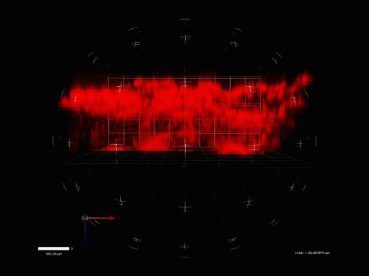
Tobacco leaf mesophylls 3D: movie showing chloroplasts recording in xyz with the resonant scanner, visualised by Volocity software (Liesche et al. unpublished)
|
Laser lines |
Beam splitter |
Emission |
Detectors |
|
355,405 nm UV,V |
fast BS |
|
|
|
458,488,496,514 nm |
tuneable AOBS |
range freely selectable |
5 PMT |
|
470-670 nm WLL |
|
|
|
|
690-1040 nm MP |
fas tBS |
2 NDD |
Details
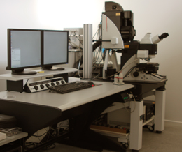
Microscope
Point Scanning Confocal and 2-photon microscope SP5-X MP UV, Leica Microsystems
Location
Frederiksberg Campus, CAB, Thorvaldsensvej 40, stair 10, 1. floor, room R164d
Typical applications
Live imaging of cells and tissues, (co-) localisation of fluorochromes and fluorescent proteins, 4D confocal imaging (xyzt), excitation and emission spectra (l2), photoactivaiton, FRET, FRAP
Consultation
Sebastian Kjeldgaard-Nintemann
Alexander Schulz
Training and problem reporting
Sebastian Kjeldgaard-Nintemann
Catherine Nielsen
Booking
Online via PPMS